What It Is For
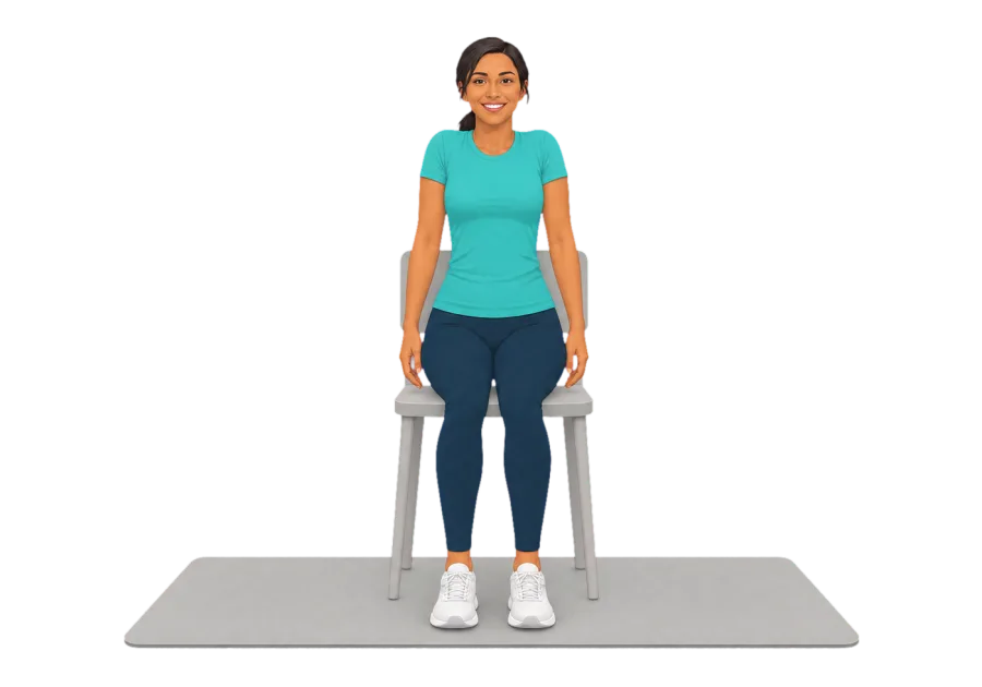
The Chin Tuck moves the head straight back over the shoulders, with the eyes level and without nodding or tipping. It is a very small movement. The result is a temporary double chin and a light stretch at the base of the skull, and it is used most by people who spend long hours with the head drifting forward towards a screen.
The NHS, writing about neck pain, advises sitting upright and gently bringing the neck back along with a roll of the shoulders.[1] The National Axial Spondyloarthritis Society teaches chin tucks with the same emphasis on level eyes, a hold of one breath and three repetitions, and notes that some tension at the back of the neck is natural.[2] Physiotherapists also use the tucked position as a starting point for other neck movements. One leaflet has patients keep the chin gently tucked while looking up, down and to the side.[3]
How to Do It
A small move that takes your head back over your shoulders after screen time. Look straight ahead and glide your chin straight back, as if making a double chin, without tipping your head. Hold, release and repeat. You should feel a light stretch at the base of your skull and the back of your neck.
- Look straight ahead
- Keep your eyes level
- Small and gentle
How It Got Its Name
Chin tuck is an informal description of the position. It may mislead, because the chin does not drop towards the chest. Clinicians prefer retraction, from the Latin for drawing back. The first printed use of chin tuck has not been traced.
The same words mean something else in speech and language therapy. There, a chin tuck or chin-down posture is a head position used during swallowing, studied in people with dysphagia.[7] This is a different movement with a different purpose, and the two should not be confused.
History
No inventor of the chin tuck is established. Physiotherapists had drawn on repeated neck movements for decades, and retraction may well predate any account of it in print. No primary source was found that fixes its first appearance.
The name most associated with it is Robin McKenzie, a New Zealand physiotherapist. His Mechanical Diagnosis and Therapy system grew from a chance discovery in 1956, and he founded the McKenzie Institute International in 1982. He published self-care books on the back in 1980 and on the neck in 1983, and a cervical and thoracic textbook in 1990. Over six million copies of the back and neck books have reportedly been sold, in seven languages.[8] Retraction is widely described as a core exercise of his neck programme, though this article could not check the 1983 text directly.
A separate line of work came from clinical research. Jull and colleagues described a new test of craniocervical flexion in a 1999 paper on neck-related headache.[9] A 2008 account by Jull, O'Leary and Falla said the test had evolved over about 15 years, in response to research on the role of the deep flexors in supporting the neck. That account also describes the use of the same action as a retraining exercise.[5] The McKenzie retraction and the Jull nod are now often lumped together as chin tucks, although they arose separately.
Muscles and Tendons
Two different actions are often called a chin tuck, and they use the neck differently. A retraction, as taught here, is a horizontal glide of the head. A craniocervical flexion is a small nod, usually practised lying down. Both work the deep neck flexors: longus colli, which lies on the front of the spine from the first cervical to the third thoracic vertebra, and longus capitis.[4][5]
These deep muscles are tested and trained at low load. A study of ten volunteers measured greater activity in the superficial sternocleidomastoid and anterior scalene during ordinary neck flexion than during craniocervical flexion, so the small action appears to be the more specific one for the deep group.[6] Light effort therefore matters, because straining brings in the larger muscles.
The stretch felt at the base of the skull comes from the upper neck flexing as the head moves back. The suboccipital muscles and the tissues at the top of the back of the neck lengthen, and the nuchal ligament is lightly tensioned. The move is mainly a short muscle contraction with a small stretch, not a sustained stretch.
Stretched
- Suboccipital muscles
- The small muscles at the base of the skull, where the upper neck flexes
- Upper fibres of the trapezius and semispinalis capitis
- A light stretch at the top of the back of the neck
Working
- Longus colli and longus capitis
- The deep neck flexors on the front of the cervical spine
- Sternocleidomastoid and anterior scalene
- Should stay fairly quiet in a gentle version
Tendons and Fascia
- Nuchal ligament
- The midline band at the back of the neck, lightly tensioned at the end of the glide
What Research Has Looked At
Much of the research concerns the lying-down nod and not the seated glide. Studies reviewed in 2008 found that people with neck pain tended to show less deep flexor activity and more superficial activity during the test.[5] A 2016 study of 32 women with chronic neck pain then recorded both muscle groups directly and found only a moderate inverse relationship between them, so high superficial activity cannot be taken as a straightforward sign of weak deep muscles.[10]
A Cochrane review update found support for neck and shoulder strengthening in chronic neck pain, and included pressure biofeedback methods of this kind among the exercises for neck-related headache.[11] These findings describe programmes in clinical groups. They do not show that a short chin tuck at a desk changes how the head is carried.
Where It Is Used Today
Chin tucks are a standard item in physiotherapy home programmes, in workplace advice and in charity guidance for people with neck stiffness.[2][3] Clinicians trained in McKenzie's system assess how a person responds to position and movement, and retraction is commonly part of that work.[8] Clinics and research laboratories use the craniocervical flexion test, with a pressure sensor behind the neck, to assess the deep flexors, usually with the person lying on the back.[5]
Variations and Related Moves
A smaller glide is the easier form. Standing with the back against a wall gives feedback about the head's position and makes the move harder to cheat. Lying on the back with a pressure cuff behind the neck turns it into the clinical craniocervical flexion exercise.[5]
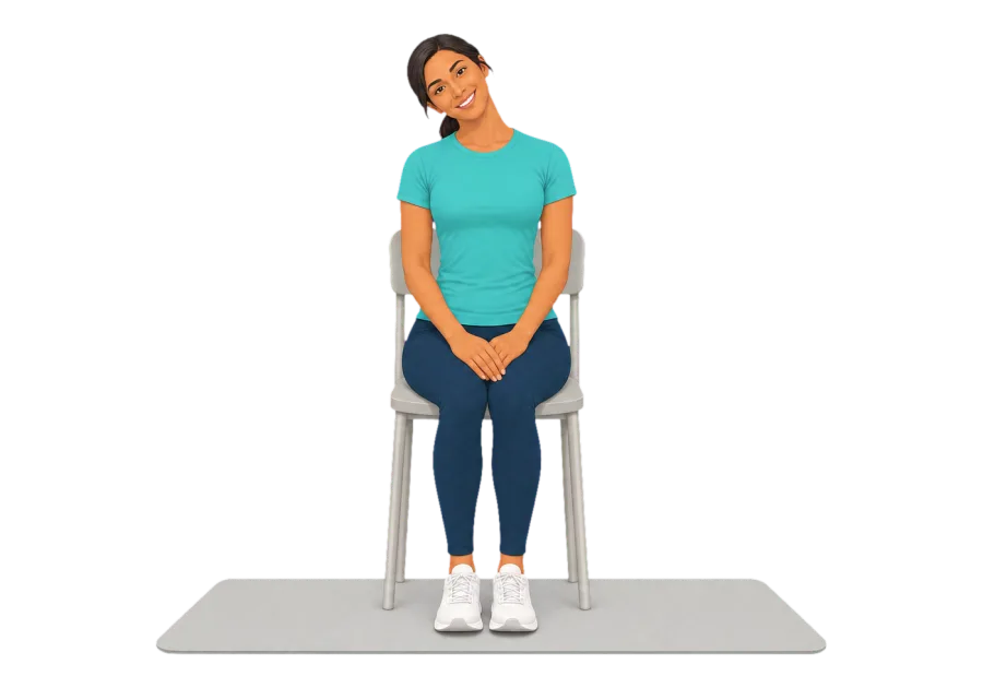
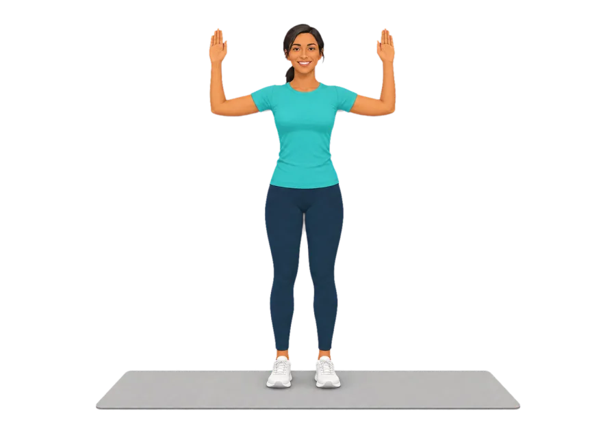
Leaflets also use the tuck as a set position, adding rotation, side bending or a look upwards from it.[3] The Neck Side Stretch and Shoulder Rolls often accompany it in desk routines.[2]
Routines With This Move
- Chair Break
- Desk Reset
- End of the Working Day
- Neck Release
- Open Shoulders
- Steady and Strong, Longer
- Upper Body Unwind
See Also
Sources
- Neck pain (opens in a new tab). NHS.
- Neck and Shoulder Stretches (opens in a new tab). National Axial Spondyloarthritis Society, 2020.
- Neck Mobility Exercises (opens in a new tab). Musculoskeletal Physiotherapy OCATS, Health Services at Wolves.
- Longus colli muscle (opens in a new tab). Wikipedia.
- Clinical assessment of the deep cervical flexor muscles: the craniocervical flexion test (opens in a new tab). Journal of Manipulative and Physiological Therapeutics, 2008.
- Muscle specificity in tests of cervical flexor muscle performance (opens in a new tab). Journal of Electromyography and Kinesiology, 2007.
- Impact of Head and Neck Posture on Swallowing Kinematics and Muscle Activation: A Systematic Review (opens in a new tab). Dysphagia, 2025.
- Robin McKenzie: the Influence of a giant (opens in a new tab). New Zealand Journal of Physiotherapy, 2013.
- Further clinical clarification of the muscle dysfunction in cervical headache (opens in a new tab). Cephalalgia, 1999.
- Does increased superficial neck flexor activity in the craniocervical flexion test reflect reduced deep flexor activity in people with neck pain? (opens in a new tab). Manual Therapy, 2016.
- Exercises for mechanical neck disorders: A Cochrane review update (opens in a new tab). Manual Therapy, 2016.
Further Reading
- Flexibility exercises (opens in a new tab). NHS.
- Exercises for the neck (opens in a new tab). Arthritis UK.
General information about this move, not medical advice. Stop if something hurts, and read our Safety page before you start. If you have a health condition, ask your doctor or physiotherapist first.